
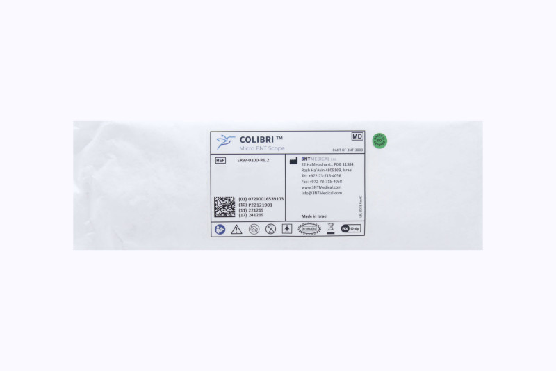
ERW-0100-R6.2
3NT Medical Colibri Micro ENT Scope
$15.00
eaches
In Stock: 2 Eaches
Expiration:
Expired
Express: Need it tomorrow? Order in next 2 hours 8 minutes and choose overnight shipping at checkout.
The item listed above is expired and may be used for educational, training, and non-clinical research purposes only. Any product information appearing below, including the product indication statement, pertains to an in-date item only.
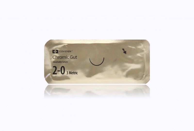
Medtronic Chromic Gut 30" V-26 taper
$4.00 packets
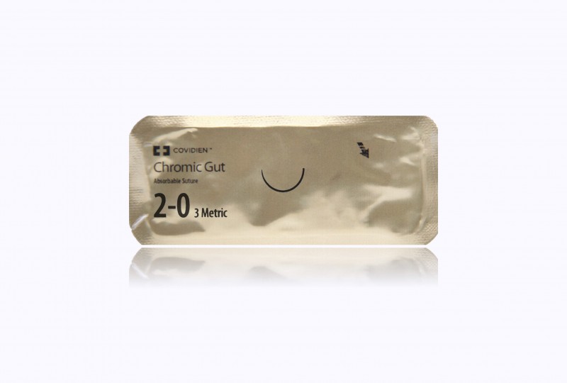
Medtronic Chromic Gut 30" GS-21 taper
$4.50 packets

Maxon green 60" GS-26 taper, looped to 30"
$8.95 packets

Medtronic Sofsilk black 30" SC-2 straight cutting
$1.00 packets
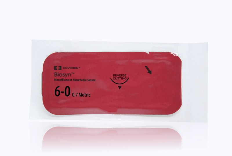
Medtronic Biosyn undyed 18" P-10 Reverse Cutting Needle
$8.00 packets
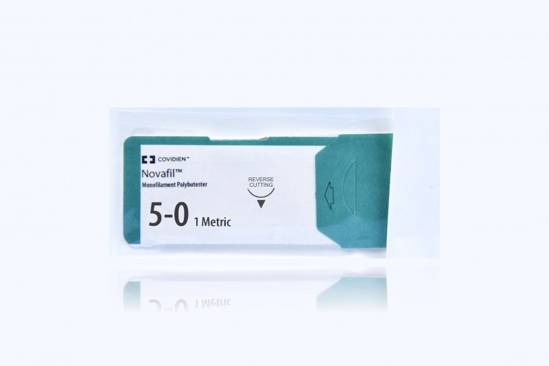
Medtronic Novafil blue 18" SBE-4 cutting
$4.00 packets
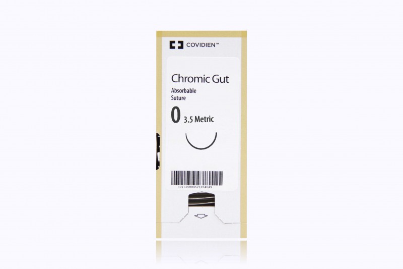
Medtronic Chromic Gut 30" GS-22 taper
$148.00 box of 36
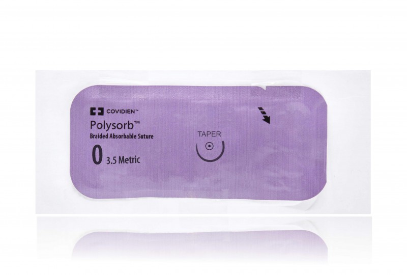
Medtronic Polysorb violet 30" GU-46 taper
$4.00 packets
Description
3NT Medical Colibri Micro ENT Scope
Colibri micro ENT scope is intended to visualize the internal cavities of the ear, airways, nose and sinus cavities during diagnostic and therapeutic endoscope procedures.
Product Code: ERW-0100-R6.2
Additional Details
- Company Name: 3NT Medical, Ltd.
- FDA Product Code: EOB
- FDA Product Code Name: Nasopharyngoscope (Flexible Or Rigid)
- GMDN Term Code: 64899
- GMDN Term Name: Rigid video otorhinolaryngoscope
- GMDN Term Definition: An endoscope with a rigid inserted portion intended for the visual examination of some or all of the ear (external, middle and inner), nasal passages, paranasal sinuses, pharynx, and/or larynx. It is inserted into the body through the appropriate natural orifice and can be used for treatment if it has a working channel. Anatomical images are transmitted to the user by a video system with an image sensor chip at the distal end of the endoscope and the images showing on a monitor (not included); it may include an irrigation/suction channel. This is a single-use device.
Additional information
| Size | 1 |
|---|---|
| Arms | N/A |
| Manufacturer | |
| Unit | eaches |
- FDA Product Code: EOB
- FDA Product Code Name: Nasopharyngoscope (flexible or rigid)
- GMDN Term Code: 64899
- GMDN Term Name: Rigid video otorhinolaryngoscope
- GMDN Term Description: An endoscope with a rigid inserted portion intended for the visual examination of some or all of the ear (external, middle and inner), nasal passages, paranasal sinuses, pharynx, and/or larynx. It is inserted into the body through the appropriate natural orifice and can be used for treatment if it has a working channel. Anatomical images are transmitted to the user by a video system with an image sensor chip at the distal end of the endoscope and the images showing on a monitor (not included); it may include an irrigation/suction channel. This is a single-use device.